Sinnecker T, Othman J, Kühl M, Mekle R, Selbig I, Niendorf T, Kunkel A, Wienecke P, Kern P, Paul F, Faiss J, Wuerfel J. 7T MRI in natalizumab-associated PML and ongoing MS disease activity: A case study. Neurol Neuroimmunol Neuroinflamm. 2015; 2(6):e171.
OBJECTIVE:To assess the ability of ultra-high-field MRI to distinguish early progressive multifocal leukoencephalopathy (PML) from multiple sclerosis (MS) lesions in a rare case of simultaneous presentation of natalizumab-associated PML and ongoing MS activity.
METHODS:Advanced neuroimaging including 1.5T, 3T, and 7T MRI with a spatial resolution of up to 0.08 mm(3) was performed.
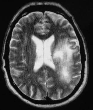
RESULTS:7T MRI differentiated between PML-related and MS-related brain damage in vivo. Ring-enhancing MS plaques displayed a central vein, whereas confluent PML lesions were preceded by punctate or milky way-like T2 lesions.
CONCLUSIONS:Given the importance of early diagnosis of treatment-associated PML, future systematic studies are warranted to assess the value of highly resolving MRI in differentiating between early PML- and MS-induced brain parenchymal lesions.
So this study looks at 7T MRI which has a resolution og 0.08mm3 so that is about 450micrometers in any one plane 0.44 (0,44 x 0,44 x 0,44 = 0.085mm3, which is about 10-11 cells in either plane so resolution of about 1000 cells and MS lesions have a central vein compared to PML. Maybe a use for T2* imaging